X-rays as a diagnostic tool in injury cases are used in car accidents, slip and falls, dog bites, medical malpractice, dental malpractice, trucking accidents and other claims. Imaging diagnostic tests are vital for establishing the cause of neck and back pain. One of the tests that your doctor may order is an X-ray of your spine if you present with such pain. Although it is not as commonly used for this purpose as CT and MRI scans, it is useful in establishing the injury that is causing you the trouble. Here are the answers from a personal injury perspective to the questions you might be having about this diagnostic tool.

What is an X-ray?
An X-ray is a diagnostic imaging technique that uses invisible electromagnetic energy beams to produce images of bones, organs and internal tissues of the human body. It is the most used imaging test now that it’s fast, simple, and widely available.
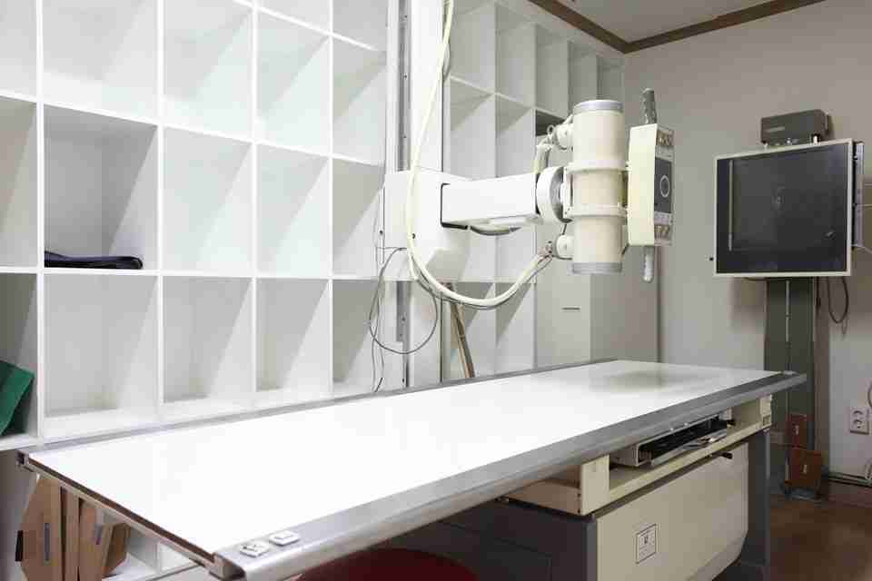
Although it may not look as sophisticated as newer imaging tests, it has evolved since the accidental discovery of X-rays in 1895. The current X-ray machine, which looks like a tube containing a large light bulb, is far advanced compared to the first one used in a radiology department in 1896.
Some of the uses of X-ray imaging include:
- Bone fractures and breaks
- Dislocation
- Cancerous and non-cancerous bone tumors
- Bone degeneration
- Infections
- Lung problems like pneumonia and lung cancer
- Heart conditions
- Breast cancer
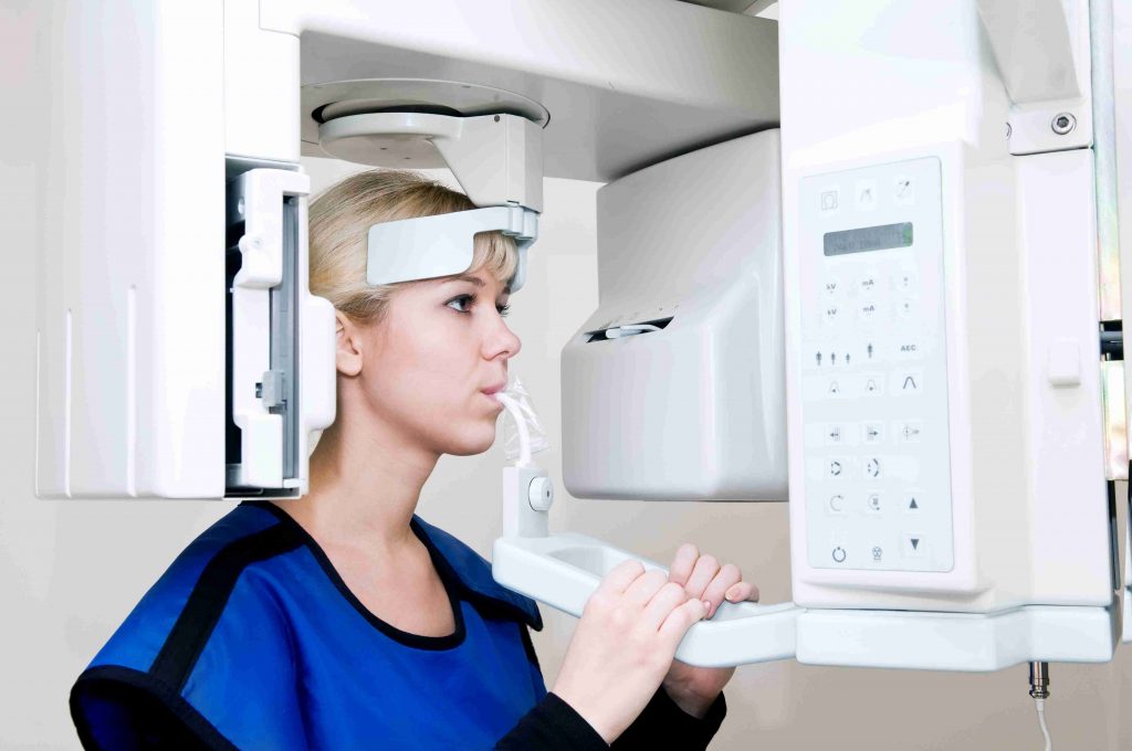
- Teeth problems
- Digestive system issues like difficulty swallowing and intestinal obstruction
How Does an X-ray Work?
You probably are wondering how exactly an X-ray machine works. Here’s what you need to know. Don’t worry; this is not going to be one of those mind-boggling physics classes.
As mentioned earlier, X-ray imaging uses invisible waves of electromagnetic energy referred to as X-rays. The rays were named X because they were unknown rays discovered accidentally during an experiment. X-rays are produced when a negatively charged electrode undergoes heating by electricity which releases electrons. The process produces energy that is directed towards a metal plate at a high velocity. Collision of the energy with atoms in the metal plate produces X-rays.
During imaging, the part being imaged is positioned between an X-ray machine that produces the X-rays and a detector. The detector has always been a photographic film held in place by a cassette. However, modern machines now use a digital X-ray sensor.
When x-rays pass through the body, there is absorption of energy at different rates depending on the body part. The denser the tissue, the more the x-rays it absorbs. Dense tissues like bone and some tumors absorb most of the X-ray energy and therefore appear light or white on the film. Soft tissues like blood, muscle, fat and skin allow most of the x-rays to pass through. Consequently, they appear dark on the X-ray image.
Given that soft tissues absorb less of the x-ray energy, they are challenging to see. That is why X-rays are mostly used for generating detailed images of the bony structure of the body. Radiographers can, however, visualize soft tissues better by injecting you with a contrasting agent if needed. However, that is rarely applicable when it comes to spine x-rays.
What Will I Experience During the X-ray?
Perhaps you are a little nervous about the whole ordeal of the imaging process. Lucky for you, the X-ray is a non-invasive and painless procedure. If you need to be injected with contrast, you will have to brave the needle prick.
It’s also a quick procedure. In fact, it is the quickest imaging test. Ten minutes is more than enough time to get the procedure done unless you need contrast imaging.
But of course, you want to know what exactly to expect during the procedure. Here is each and every step of the imaging process.
Preparation for an X-ray
There is really nothing much that you need to do in the preparation period. You don’t have to stop eating or drinking before the procedure. Additionally, sedation is not necessary unless you are in so much pain that staying calm is difficult.
If you need a contrast agent, you will be told to stop eating, drinking or taking certain medications for a few hours before the imaging.
Your radiographer may want to know if you are pregnant. If you are not sure, a pregnancy test may be necessary due to the potential harm the X-rays may have on your baby.
Loose and comfortable clothes are perfect for imaging. It’s okay to still show up with your picture-perfect outfit; however, the radiographer will ask you to change into a hospital gown during the imaging. You will also have to remove jewelry, hairpins, hearing aids, eyeglasses and other metallic objects that can interfere with the imaging.
The X-ray Procedure
In the imaging of your spine, your radiographer will ask you to stand against a flat surface or lie on a table. Once you are in the proper position, they will carefully aim the x-ray machine at your spine. A lead shield may be used to cover the parts that are not being imaged. The radiographer will ask you to remain still during the imaging and then move to the adjacent room or behind a screen where the machine is operated.
You will feel nothing as the X-ray is done. The procedure is fast as each image lasts for a fraction of a second. A spine X-ray necessitates taking images at different angles. The radiographer will therefore move the machine between images to provide as much detail as possible.
As mentioned earlier, the procedure takes under ten minutes. You can go home shortly afterwards as it has no side effects. It’s a non-invasive procedure, and therefore, you can return to your normal activities straight away. However, you may need to stick around for some time in case contrast was used. The radiographer will monitor you for a short while to ensure you don’t have any side effects. In case you develop some, they are usually temporary and will disappear in a short while.
Interpretation of the X-ray Images
The X-ray images obtained are either written on a CD or developed from a photographic film. Images written on a CD can be viewed on any computer screen. Besides these images or films, your doctor will also require the interpretation of these images by a radiologist.
A radiologist is a doctor specifically trained to supervise and interpret radiological examinations. They will examine the images and compile a report. They may discuss the results with you on the same day or send the report to your doctor, who will discuss the findings with you later.
Advantages and Disadvantages of X-ray Imaging
If you have been wondering whether the X-ray is really the best procedure for you, here are the pros and cons you may need to know.
Advantages of X-ray Imaging
- Simple and fast
- Non-invasive and painless
- Cheaper than other procedures like CT and MRI
- Less radiation exposure as compared to CT scans
- Widely available
Disadvantages of X-ray Imaging
- Cannot generate 3D images, unlike X-ray and CT
- Less detailed images
- Exposure to ionizing radiation
Risks of X-ray Use to Your Health
As for any other imaging diagnostic test, the benefits must outweigh the risk. X-ray imaging has two risks that you need to understand:
- Ionizing radiation
- Contrast agent
Ionizing Radiation
According to studies, X-rays are a form of radiation that can potentially result in damage to cells in the human body and subsequent development of cancer. However, the risk is really small. Here are the numbers to put everything into perspective according to FDA.
Exposure to 10 millisieverts (mSv) from an X-ray or CT increases your risk of dying from cancer by 0.05%. A single X-ray exposes you to 0.28mSv; therefore, you will need about 36 x-rays in your lifetime to increase your cancer risk by 0.05%. X-rays are way safer than CT scans which expose you to 6.3mSv per scan, thus increasing your risk by 0.05% after only two scans. As long as you are keeping the number of X-rays low, your risk is low.
Contrast Agent
There is also some risk associated with the use of contrast agents. Although your spine X-ray will not require a contrasting agent, you need to understand what risk comes with it in case it’s used. You may experience some side effects. Inform your radiographer if you have any allergies, as the agent may trigger an allergic reaction. It’s advisable not to use the contrast if you have kidney disease as kidneys are involved in clearing the agent from your body.
Uses of X-ray Imaging in Neck and Back Pain
An X-ray scan is an excellent tool for evaluating the cervical, thoracic, lumbar, sacral and coccygeal regions of your spine. The main uses of the X-ray in the management of your neck and back pain include:
- Detection of the injuries causing your pain
- Planning of your treatment
- Assessing the efficiency of the treatment
X-rays are instrumental in the diagnosis and treatment sustained from construction accidents, car wrecks, slip and falls and trucking accidents.
Symptoms of Neck and Back Injury
How do you know that the neck and back pain is due to your accident? You can lookout for the following symptoms besides pain that point towards neck and back injury after a car wreck:
Immediate Symptoms
- Weakness or paralysis of your fingers or toes
- Altered sensation in your arms or legs, for instance, tingling or burning sensations and numbness
- Headaches
- Loss of bladder or bowel control
- Muscle spasms
- Sexual dysfunction
Delayed Symptoms
- Headaches
- Tingling or burning sensation or numbness
- Weakness in your hands or legs
Neck and Back Injuries Detectable on X-ray
Despite its lack of detail that minimizes its usefulness in spine injuries, X-rays are still used. Most specialists refer to them as the ‘quick and dirty’ way to assess a spine for injuries and abnormalities especially in emergency rooms for car accidents, trucking accidents, construction and slip and fall accidents. They come in handy as they are quick and readily available.
An X-ray can reveal some helpful information and is necessary even if you need more sophisticated imaging tests. For instance, surveillance radiographs are taken after accidents to rule out any injuries requiring immediate attention.
The injuries of the spine detectable on X-ray include:
- Vertebral fractures
- Disc herniation
- Connective tissue injuries
Vertebral Fractures
Perhaps the commonest use of X-rays in assessing spinal injuries is in the detection of fractures of the vertebral bones. Fractures in the spine result in displacement of the bones that can compress on nerves and result in irritation felt as pain. Additionally, a fractured vertebra is unstable and may injure the spinal cord that may cause paralysis and other problems.
A plain X-ray can help detect these fractures. However, hairline fractures and non-displaced ones are challenging to detect using an X-ray. You may require advanced modalities for better visualization of these injuries.
Disc Herniation
Excessive compressive force on the spongy discs between your vertebral bones can result in their damage and outward displacement. The displacement can irritate the nearby nerves through compression, which you will perceive as pain.
Imaging using an X-ray machine can reveal some of these injuries. It may not show the minute details about the disc injury; however, it can point towards what is causing the neck and back pain.
Connective Tissue Injuries
When the best possible technique is used, X-rays can show injuries in the connective tissues of the spine that might be the reason for your pain. Injuries of the ligaments in the spine, for instance, can be picked up on a plain radiograph. However, it will require a very keen eye on the part of the radiologist to identify these injuries.
If the imaging results are inconclusive, you may need other tests like CT scanning and MRI. The MRI scan is especially best suited for the assessment of soft tissues. Your radiologist or doctor may recommend these follow-up images to understand better the reason behind your neck is and back pain.
Conclusion
Hopefully, you now understand why your doctor may have ordered that spine X-ray. Although it is not as popular in imaging the neck and back as it is in imaging other parts of the body, it is still useful. Doing the X-ray will bring you a step closer to finding a solution to your nagging neck and back pain.
CALL TODAY FOR A FREE CONSULTATION WITH OUR ACCIDENT ATTORNEY 713-572-6446
This page is for informational purposes provided by the personal injury law firm of Jerome O. Fjeld, PLLC has served injury victims in Texas for nearly two decades. Our firm’s primary office is in Houston, TX with offices in Austin and Victoria. Our firm’s focus is personal injury and we help victims when someone is injured by the negligence or carelessness of another in the following types of cases:
- dog bites and dog attacks,
- trucking accidents,
- car accidents,
- construction accidents,
- uber accidents,
- boating accidents,
- work accidents,
- slip and falls,
- bike accidents,
- accidente de carro and
- other cases when someone is seriously injured by the negligence or carelessness of another.